Smart Lapidus℠

The SMART Bunion technology was designed to give your empower your surgeon to generate reproducible outcomes tailored to you.
What is SMART Bun-Yo-Matic℠?
We understand there is not a one-size fits all approach for bunion treatment. Smart Bunion is intended to assist in the pre-operative planning of your unique bunion deformity. Your scans are loaded into our software and AI-driven algorithms model your food to export a surgical plan specifically for your correction.
SMART Bun-Yo-Matic is the first bunion device on the market that allows the surgeon to see the anatomy of your foot in 3D using only standard 2D x-rays.
We want to give the surgeon the most intelligent, advanced tools to accelerate and simplify your procedure.
There are many other bunion products in the market, but this is the only custom designed specifically for you. You need an experienced foot and ankle surgeon to correct your bunion in a personalized and precise way for you versus a generic approach. The most successful foot and ankle surgeons seek out the greatest control possible in their corrective surgery and all the latest procedures. The Smart bunion is designed to give your surgeon the greatest control possible ensuring the replicable results you deserve.
Your AI Driven Second Opinion
Get reproducible correction with minimal joint disruption
We understand you are in pain and that you want to get back on your feet as quickly as possible after surgery. We also understand that your surgeon is the hero and wants the same thing for you.
We understand you are in pain and that you want to get back on your feet as quickly as possible after surgery. We also understand that your surgeon is the hero and wants the same thing for you.
With the Smart Bunion technology, your surgeon is going to design a custom solution for you, taking into consideration your unique needs.
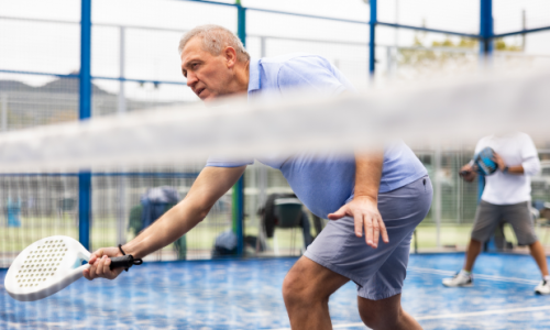
Engage in regular actives faster
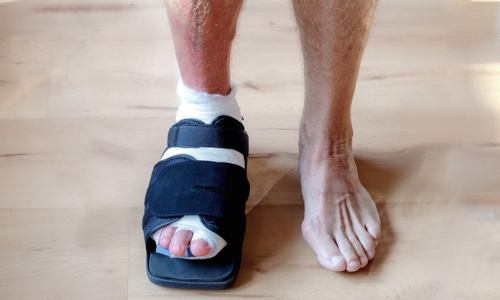
Day of Surgery
Following a Lapidus procedure, the patient needs to be non-weight bearing for 4 weeks. Progressive weight bearing in a boot start after 4 weeks.
Immobilization starts first 2 weeks in a splint (half-cast) followed by 6 weeks in a tall boot.”
4 to 6 Weeks
At 6 weeks, patient is full weight bearing in a boot and can transition out of the boot into rocker-bottom, stiff-sole shoe wear.”

6 to 8 Weeks
Patients can resume light activities once they are full weight bearing in a boot. At that point, they can do stationary bike, elliptical and low impact activities.
8 Weeks and Beyond
Activities improve even more once you are out of the boot into a surgical shoe at 8 weeks. Higher impact activities such as walking as an exercise and jogging can be resumed after 3 months. Contact sports or full activity are not advised before 4 months postoperatively.
Day of Surgery
Following a Lapidus procedure, the patient needs to be non-weight bearing for 4 weeks. Progressive weight bearing in a boot start after 4 weeks.
Immobilization starts first 2 weeks in a splint (half-cast) followed by 6 weeks in a tall boot.”
4 to 6 Weeks
At 6 weeks, patient is full weight bearing in a boot and can transition out of the boot into rocker-bottom, stiff-sole shoe wear.”
6 to 8 Weeks
Patients can resume light activities once they are full weight bearing in a boot. At that point, they can do stationary bike, elliptical and low impact activities.
8 Weeks and Beyond
Activities improve even more once you are out of the boot into a surgical shoe at 8 weeks. Higher impact activities such as walking as an exercise and jogging can be resumed after 3 months. Contact sports or full activity are not advised before 4 months postoperatively.
Animation
AI-driven algorithms and statistical shape modeling work with surgeon input to minimize bone resection and help restore natural foot function

Paragon 28® operates under the following principles:
Inclusion and respect of individual surgeon’s preferences, creative innovation, high-quality, cost-effective implants, and a strong belief that through research and innovation, we can create new and improved solutions to the challenges faced by foot and ankle specialists.
Recovery
Typically, MIS surgery allows for faster recovery and weightbearing. Depending on your surgeons’ treatment, this may vary.
Frequently Asked Questions
Why was SMART Lapidus ℠ technology developed?
SMART Lapidus SM was developed because each patient’s anatomy is unique. Surgeons have different comfort levels with many procedures and this technology enables a surgeon to correct a bunion deformity precisely to each patient.
What do you mean by ‘SMART’?
The SMART Lapidus SM software is intended to assist in the pre-operative planning of hallux valgus deformities. A weight bearing CT scan of the patients’ foot is imported into our software. AI-driven algorithms and statistical shape modeling work with surgeon input to export a surgical plan for correction. Cutting guides are then designed to precisely correct the bunion.
How is this different from other bunion correction procedures on the market?
Other bunion products have been used to correct deformities for years as standard of care. But as healthcare treatments advance, we see the need to treat everyone uniquely to their anatomy.
SMART Lapidus SM is an advancement in instrumentation such that the surgeon is not making freehand cuts to estimate correction, but the system allows for calculated bone cuts.
What is the healing time and how long will my mobility be limited?
All restrictions related to shoes are at the discretion of your surgeon. Patients may return to normal footwear in as little as 4-6 weeks.
When can I return to normal activities?
All restrictions related to activities are at the discretion of your surgeon. Patients may return to normal footwear in as few as 4-6 weeks.
Is this procedure covered by insurance?
Coverage is dependent upon the provider and your insurance. The SMART Lapidus SM procedure is a bunion correction procedure, which is a common occurrence for insurers.
Where do I find a surgeon that performs this procedure?
Surgeons are being trained every week on this novel procedure. To find a surgeon near you, please utilize our surgeon finder tool.
What pre-existing health conditions would keep me from having the SMART Lapidus ℠ procedure?
The Paragon 28 SMART Lapidus SM system is not designed or sold for any use except as indicated. Use of the SMART Lapidus SM system is contraindicated in the following situations:
- Patient has an active infection.
- Significant changes to the patient’s anatomy have occurred since the medical scan used for product definition was obtained.
- CT scan date is greater than 6 months from the patient’s surgery date.
- Patient has open epiphyseal growth plates.
- Indications not included in the INDICATIONS FOR USE
Important Risk Info
Only a surgeon can tell if the Phantom® Intramedullary Nail, Phantom® MIS Procedure and/or Precision® MIS Bunion System is right for you. There are potential risks, and recovery takes time. Potential risks include but are not limited to infection, discomfort, or swelling due to balancing and introduction of the implant, loosening of the implant, and loss of correction. Refer to full list of warnings precautions, and contraindications within the Phantom® Small Bone Intramedullary Nail and Precision® MIS Bunion System Instructions for Use at https://paragon28.com/ifus/
Surgeon Finder Disclaimer
The surgeon information listed in the Surgeon Finder is provided for informational purposes only and does not represent an endorsement or warranty of any particular surgeon. The database does not include an exhaustive list of all surgeons within a particular geographic area or all surgeons who use/have used a Paragon 28® product. Only those who have expressly subscribed to be listed on the site and are confirmed to be either Paragon 28® trained and/or experienced are included. These are the only criteria for inclusion. Paragon 28® does not pay a fee or any other type of remuneration for participation. Choice of surgeon should be solely based upon a patient’s own investigation of a particular surgeon’s training, education, experience and reputation.
For the contraindications, potential complications and adverse reactions, warnings and precautions associated with this device, please refer to the device specific instructions for use here.
Sources
Lee M, Walsh J, Smith MM, Ling J, Wines A, Lam P. Hallux valgus correction comparing percutaneous chevron/Akin (PECA) and open scarf/Akin osteotomies. Foot Ankle Int. 2017;38(8):838-846. doi:10.1177/1071100717704941
Maffulli N, Longo UG, Oliva F, Denaro V, Coppola C. Bosch osteotomy and scarf osteotomy for hallux valgus correction. Orthop Clin North Am. 2009;40(4):515-524. doi:10.1016/j.ocl.2009.06.003
Blitz NM. Current concepts in minimally invasive bunion surgery. Podiatry Today. 2019;32(2):28-34.
Lam P, Lee M, Xing J, Di Nallo M. Percutaneous surgery for mild to moderate hallux valgus. Foot Ankle Clin. 2016;21(3):459-477. doi:10.1016/j.fcl.2016.04.001 (76% Smaller Incisions)
Nix S, Smith M, Vicenzino B. Prevalence of hallux valgus in the general population: a systematic review and meta-analysis. J Foot Ankle Res. 2010;3:21. Published 2010 Sep 27. doi:10.1186/1757-1146-3-21
DiDomenico LA, Wargo-Dorsey M. Lapidus Bunionectomy: First Metatarsal Cuneiform Arthrodesis. McGlamrys Comprehensive Textbook of Foot and Ankle Surgery. 4th; p. 322-330.
Yamamoto Y, Yamaguchi S, Muramatsu Y, et al. Quality of Life in Patients With Untreated and Symptomatic Hallux Valgus. Foot Ankle Int. 2016;37(11):1171-1177. doi:10.1177/1071100716655433
Gribbin CK, Ellis SJ, Nguyen J, Williamson E, Cody EA. Relationship of Radiographic and Clinical Parameters With Hallux Valgus and Second Ray Pathology. Foot Ankle Int. 2017;38(1):14-19. doi:10.1177/1071100716666562
Cronin, S., Conti, M., Williams, N., & Ellis, S. J. (2020). Relationship Between Demographic and Radiographic Characteristics and Second Ray Pathology in Hallux Valgus Patients. Foot & Ankle Orthopaedics. https://journals.sagepub.com/doi/full/10.1177/2473011420909088
Lai, M. C., Rikhraj, I. S., Woo, Y. L., Yeo, W., Ng, Y. C. S., & Koo, K. (2018). Clinical and Radiological Outcomes Comparing Percutaneous Chevron-Akin Osteotomies vs Open Scarf-Akin Osteotomies for Hallux Valgus. Foot & Ankle International, 39(3), 311–317. https://journals.sagepub.com/doi/10.1177/1071100717745282
Peterson KS, McAlister JE, Hyer CF, Thompson J. Symptomatic Hardware Removal After First Tarsometatarsal Arthrodesis. J Foot Ankle Surg. 2016;55(1):55-59.doi:10.1053/j.jfas.2015.06.001
Prieto-Diaz, C., Anderle, M. R., Brinker, L. Z., Allard, R., & Leasure, J. (2019). Biomechanical Comparison of First Tarsometatarsal Arthrodesis Constructs Over Prolonged Cyclic Testing. Foot & Ankle Orthopaedics. https://journals.sagepub.com/doi/10.1177/2473011419892240